Immuno-Oncology · Insight
When the brake doesn't lift: how anti-PD-1 therapy affects the antibody response
PD-1 blockade reinvigorates killer T cells — but what does it do to the antibody side of immunity?
A response that protects us — and can also harm us
Our immune system has two great arms. One is cellular, built around the "killer" T cells that destroy infected or cancerous cells. The other is humoral: the production of antibodies by B lymphocytes. These antibodies are remarkable molecules. They neutralize viruses, flag bacteria for destruction, and form the basis of the protection we gain from classical viral vaccines. Yet the very same machinery, when it turns against the body's own tissues, drives autoimmune disease — and, in transplant recipients, fuels the antibody-mediated rejection of a life-saving graft. The humoral response is therefore a double-edged sword: essential to our defense, but potentially deleterious when misdirected.
A decade of immunotherapy — and a surprising blind spot
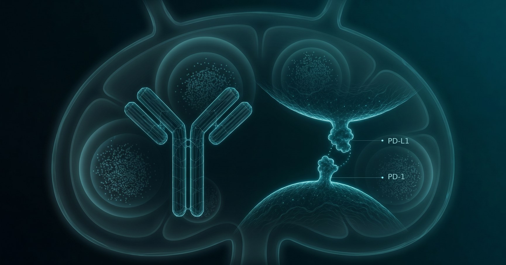
For more than ten years, immune checkpoint inhibitors targeting the PD-1/PD-L1 axis have transformed the treatment of many cancers, from melanoma to non-small cell lung cancer (NSCLC). We now understand their dominant mechanism well: by blocking the inhibitory PD-1 receptor, these antibodies "release the brake" on exhausted cytotoxic CD8+ T cells, reinvigorating the antitumor response.
What is striking — and far less appreciated — is how little we know about what these drugs do to the humoral response, whether antitumor, autoimmune, or antiviral. For a class of medicines given to so many patients for so long, this is a genuine knowledge gap.
It is made all the more surprising by a second observation: the preclinical animal models routinely used to characterize new molecules before they reach humans are largely silent on this question.
What theory predicts: an amplified antibody response
Based on what those animal models do tell us, the expectation is clear. PD-1 is not confined to cytotoxic CD8+ T cells: it is highly expressed on follicular helper T cells (Tfh), the CD4+ cells that orchestrate B-cell maturation in germinal centers, as well as on B cells themselves. Since PD-1 delivers an inhibitory signal when it engages PD-L1, blocking it should, in principle, lift a brake on antibody production as observed in experimental models with antigen-specific antibodies1. On the contrary, when PD-1 signaling is disrupted from the initial steps of B-cell development, B-cell maturation and antibody responses are impaired2, illustrating the complexity of PD-1/PD-L1 regulation of humoral immunity. However, since patients are not deficient in PD-1 but rather have it transiently blocked, one would expect anti-PD-1 treatment to enhance, rather than impair, the humoral response.
What the human data actually show: weak, variable, hard to interpret
A handful of human studies have tried to test this prediction in antiviral or antitumor settings — and the picture is murky. The effects reported are generally modest, short-lived, variable, or limited to the biochemical features of the antibodies rather than their quantity. After influenza vaccination, anti-PD-1 reshaped circulating Tfh and B-cell dynamics without translating into a clearly stronger protective response3,4. In lung cancer patients vaccinated against SARS-CoV-2, PD-1 monotherapy did not significantly alter antibody titers5.
The problem is methodological as much as biological. Most studies measure pre-existing, sometimes non-specific responses, in small numbers of patients, and compare them to control groups that differ from the treated patients in age or clinical situation. The question has remained, frankly, open.
A rare clinical setting to address the question more directly
This is where our recent work provides a clearer answer. In a particular and well-controlled clinical setting, we studied the anti-HLA antibody response in patients with the same cancer, treated with or without anti-PD-16.
Why is this model so informative? Several reasons converge:
- The anti-HLA response obeys the same rules as many classical antibody responses — it is strictly dependent on follicular helper CD4+ T cells, which express PD-1 abundantly. It is therefore a faithful readout of the Tfh–B-cell axis that anti-PD-1 is predicted to disinhibit.
- We measured a genuine de novo, specific response — directed against at least four distinct HLA antigenic targets — rather than a re-stimulated pre-existing one, with a follow-up extending over two years.
- Critically, both arms — vaccine alone and vaccine plus anti-PD-1 — were composed of NSCLC patients drawn from the same clinical trial (PDC-LUNG-01, NCT03970746) and exposed to the same immunogenic stimulus: an allogeneic plasmacytoid dendritic cell vaccine (PDC*lung01). The control group was cancer patients, not healthy volunteers.
- Finally, rather than describing only the biochemical characteristics of the antibodies, we tested their function — their capacity to kill target cells through complement-dependent cytotoxicity (CDC).
The result: the brake stays on
Across 72 patients, the findings are unambiguous. Using a highly specific and sensitive antibody-detection technique, we found that about half of the patients developed anti-HLA antibodies — reaching high levels — with the response driven primarily by vaccine dose. The kinetics were consistent — antibodies appearing after the sixth injection, peaking at one month, then declining over two years, with class II preceding class I.
And anti-PD-1 changed none of it. PD-1 blockade did not increase the magnitude, the kinetics, the targets, or the cytotoxic potential of this de novo humoral response. Even when we made the conditions deliberately favorable — neutralizing the complement-regulatory proteins that protect target cells — the antibodies from anti-PD-1-treated patients were no more cytotoxic than those from patients given the vaccine alone6.
What this means
This is a genuine paradox, and an instructive one. In the very same patients, PD-1 blockade delivers clinical benefit by unleashing tumor-specific CD8+ cytotoxic immunity, while leaving the allogeneic humoral response untouched. The checkpoint is not a single master switch that uniformly "disinhibits" the immune system; its regulatory role in humoral immunity is more nuanced and more compartmentalized than the textbook model predicts.
The practical implications matter. Anti-PD-1 agents are increasingly used in solid-organ transplant recipients who develop cancer, where the fear is that lifting PD-1 might unleash donor-specific antibodies and precipitate graft rejection. Our data suggest that, at least for the generation and effector function of alloantibodies, this concern may be more limited than feared — graft rejection under checkpoint blockade is more likely driven by T-cell-mediated mechanisms than by an amplified antibody attack.
More broadly, the work is a reminder that humoral immunity remains the under-explored frontier of checkpoint inhibition — and that carefully designed human models, with matched patient cohorts and functional readouts, are what will turn assumptions into knowledge. As the field increasingly links antibody responses to immunotherapy outcomes, understanding when the brake lifts — and when it does not — will be central to designing the next generation of combination therapies and cancer vaccines.
References
- Zhang M, Xia L, Yang Y, et al. PD-1 blockade augments humoral immunity through ICOS-mediated CD4+ T cell instruction. Int Immunopharmacol. 2019;66:127–138. doi:10.1016/j.intimp.2018.10.045
- Ogishi M, Kitaoka K, Good-Jacobson KL, et al. Impaired development of memory B cells and antibody responses in humans and mice deficient in PD-1 signaling. Immunity. 2024;57:2790–2807.e15. doi:10.1016/j.immuni.2024.10.014
- Herati RS, Knorr DA, Vella LA, et al. PD-1 directed immunotherapy alters Tfh and humoral immune responses to seasonal influenza vaccine. Nat Immunol. 2022;23:1183–1192. doi:10.1038/s41590-022-01274-3
- Läubli H, Balmelli C, Kaufmann L, et al. Influenza vaccination of cancer patients during PD-1 blockade induces serological protection but may raise the risk for immune-related adverse events. J Immunother Cancer. 2018;6:40. doi:10.1186/s40425-018-0353-7
- Valanparambil RM, Carlisle J, Linderman SL, et al. Antibody response to COVID-19 mRNA vaccine in patients with lung cancer after primary immunization and booster: reactivity to the SARS-CoV-2 WT virus and Omicron variant. J Clin Oncol. 2022;40:3808–3816. doi:10.1200/JCO.21.02986
- Planel S, Vayssière G, Maggipinto G, et al. PD-1 blockade does not enhance alloimmunization after allogeneic dendritic cell vaccination in cancer patients. Front Immunol. 2026;17:1763434. doi:10.3389/fimmu.2026.1763434 · PMID 41859082